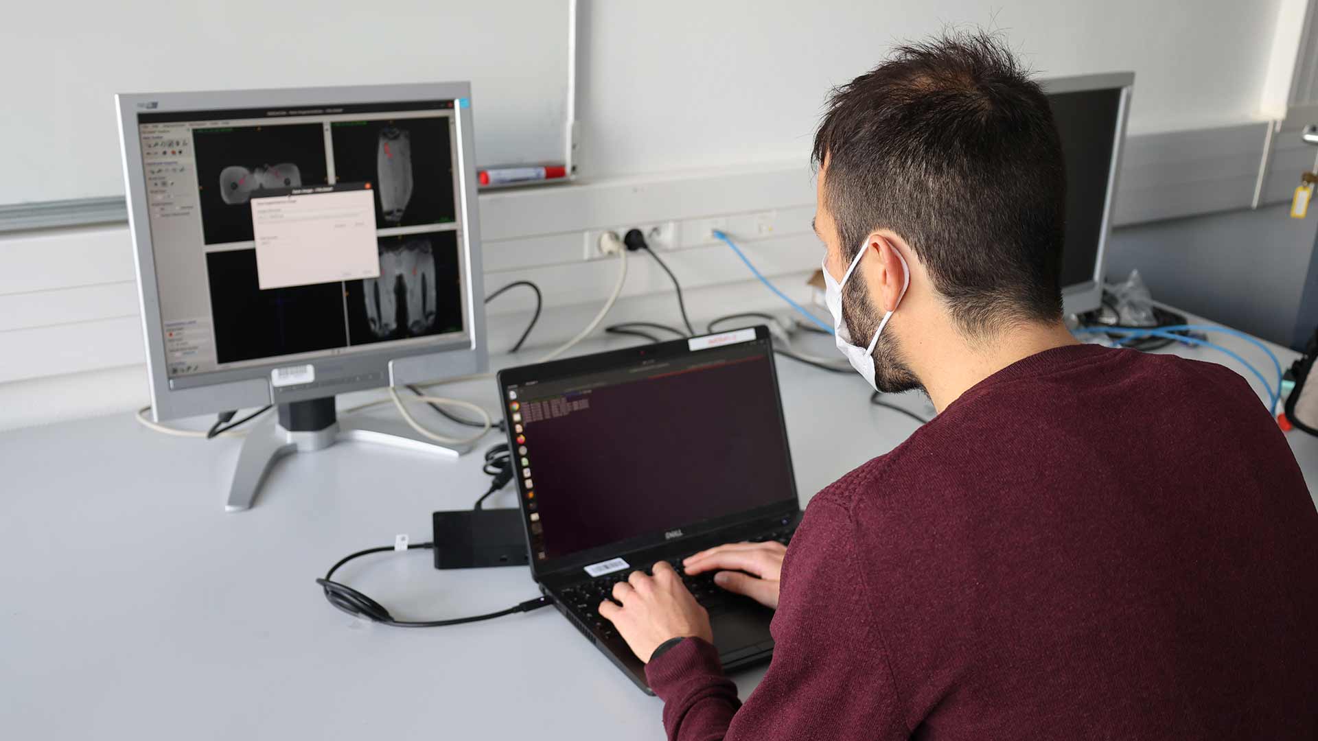
For more than 5 years, Ali Mansour, a professor at ENSTA Bretagne/ Lab-STICC, has been working alongside doctors at the Brest University Hospital (CHRU) to determine new medical image processing methods. He and his team use automatic classification and artificial intelligence techniques to improve the interpretation of ultrasound, elastography and MRI images. The aim is to better identify and characterize certain pathologies.
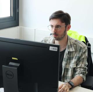
© Thibaud Berthomier / ENSTA Bretagne - Lab-STICC
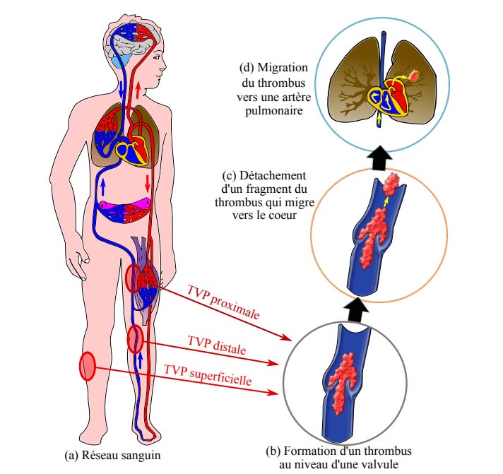
Early work on deep vein thrombosis, more commonly known as phlebitis.
This disease, the main complication of which is pulmonary embolism, is known to be multifactorial. It combines genetic factors and acquired risk factors (cancer, immobilization, surgery, pregnancy).
Between 2017 and 2019, as part of his PhD thesis (1) at ENSTA Bretagne, Thibaud Berthomier sought to identify and characterize thromboses by analyzing acoustic images (ultrasound and elastography) in order to determine the structure of blood clots known as "thrombus" and to try to deduce the factor or factors responsible for their formation.
One of the difficulties encountered was image quality: depending on the type of equipment used, the operator, the sensor position, etc., the differences encountered made the characterization of images more complex. In order to better calibrate them and thus improve processing quality, Thibaud's thesis work allowed the data acquisition protocol to be developed and better standardized.
First conclusive results for Gougerot-Sjörden's syndrome (salivary glands)
In parallel, the image pre-processing method developed by Thibaud was tested on ultrasounds and elastographies of salivary glands. The first results were conclusive for Gougerot-Sjördren's disease. They will be covered in a scientific publication.
The contribution of artificial intelligence
Since October 2020, Aurélien Olivier has been pursuing this work as part of a second thesis (2) at ENSTA Bretagne (Lab-STICC laboratory) in cooperation with Dr. Clément Hoffmann (thesis work as well).
His research on blood clot detection through deep image analysis incorporates data from an imaging database that includes ultrasounds, MRI, CT scans and other scans.
The anonymous data, acquired by several French and Dutch hospitals, combine images and medical information (patient risk factors). The work therefore involves classifying images and identifying correlations between a thrombus structure and the corresponding clinical phenotype.
In the long term, the aim is to provide practitioners with additional information on risk factors: information useful for diagnosis but not detectable by the human eye.
It is a real pleasure to work with the Brest University Hospital's teams on these innovative and meaningful subjects. The first encouraging results have opened up new research perspectives in the medical imaging field. They offer the prospect of more accurate imaging results incorporating augmented data that are useful for practitioners.
(1) Identification and characterization of venous thrombus by B-mode ultrasound imaging coupled with elastography. Thesis by Thibaud Berthomier, defended on 13/11/18 at ENSTA Bretagne and financed by the Brest University Hospital (CHRU) and the Brittany Region (Région Bretagne). Link: https://tel.archives-ouvertes.fr/tel-02366284/document
(2) Statistical methods and deep learning for the characterization of deep vein thrombosis in ultrasound and elastography. Aurélien OLIVIER's thesis, started in October 2020 at ENSTA Bretagne, financed by the Brest University Hospital (CHRU) and Brest Métropole.
Other scientific publications on this topic:
T. Berthomier, A. Mansour, L. Bressollette, F. Le Roy and D. Mottier, "Deep venous thrombus characterization: ultrasonography, elastography and scattering operator", Advances in Science, Technology and Engineering Systems Journal Vol. 2, Volume 2, Issue 3, Page No 48-59 (2017).
T. Berthomier, A. Mansour, L. Bressollette, F. Le Roy, D. Mottier, Venous blood clot structure characterization using scattering operator, in the 2nd International Conference on Frontiers of Signal Processing (ICFSP 2016), Warsaw, Poland, October 15-17, 2016.
T. Berthomier, A. Mansour, L. Bressollette, F. Le Roy, D. Mottier, Venous blood thrombosis: database creation, image preprossesing, in the 2nd International Conference on Frontiers of Signal Processing (ICFSP 2016), Warsaw, Poland, October 15-17, 2016.
T. Berthomier, A. Mansour, L. Bressollette, F. Le Roy, D. Mottier L. Frechier, B. Hermenault, Scattering Operator and Spectral Clustering for Ultrasound Images: Application on Deep Venous Thrombi, in the 19th International Conference on Biomedical Signal and Image Analysis (ICBSIA 2017), Paris, France, November 20-21, 2017
T. Berthomier, A. Mansour, L. Bressollette, D. Mottier, F. Le Roy, B. Hermenault, C. Hoffmann, L. Frechier, Unsupervised clustering using High Order Statistics Analysis for Ultrasound Images of Thrombi, In IEEE International Conference on Bioinformatics and Biomedicine (BIBM 2018), Madrid, Spain, December 3-6, 2018.
contact
Ali Mansour
Professor
IT Departement
Lab-STICC laboratory / T2I3 Departement / SI Team
+33 (0)2 98 34 87 88